MIT Developed Novel Pathogen System Based on Janus Emulsions
 Rapid methods for pathogen testing have been gaining acceptance in the food industry. Recent advances in technology result in faster detection and identification of pathogens, more convenient, more sensitive, more reproducible, and more specific than conventional methods. Many new methods are available involving antibody-based assays, genetic amplification methods, and newer sensor development methods.
However, the industry is always looking for faster, simpler and cost effective new methods. An article in ACS Central Science describes the work of researchers at Massachusetts Institute of Technology (MIT) that are developing a new method for pathogen detection, utilizing Janus emulsions. The team is lead by Timothy Swager and Qifan Zhangis the lead author.
The test is based on the analysis of liquid droplets (Janus droplets, or Janus emulsion) that are powerful liquid phase sensing particles. These droplets are formed from two equally sized hemispheres. One half is composed of a fluorocarbon and the other from a hydrocarbon.
The fluorocarbon is denser than the hydrocarbon when droplets sit on a surface, therefore the fluorocarbon orients to the bottom. When the different hemispheres are functionalized to have orthogonal physical and biochemical properties, they can be used as sensors. Consequently Janus particles with covalently modified surfaces have been used for sensing applications.
From above the droplets are transparent but when viewed sideways they appear opaque. This property relates to the way that light passes through the droplet, and it is the path of light that can be adapted to make the sensor.
Building on this the scientists at MIT developed a surfactant molecule that contains mannose sugar to form the top half of the droplet surface. These molecules are capable of binding to a protein called lectin. Lectin is a protein that can bind specifically to certain sugars and cause agglutination of particular cells, and it is found on the surface of strains of E. coli.
The emulsion assay uses the carbohydrate surfactant molecule, which self-assembles at the droplet surfaces during the emulsification process. Therefore, no further element is required for bacterial recognition. These changes in the alignment of the Janus droplets are used for the detection of analytes. The droplets are capable of binding to specific bacterial proteins. The mannose surfactant functionalized emulsion assay described in this work was designed specifically for E. coli as a model system.
Whenever E. coli is present the droplets attach to the Lectin proteins. This causes the droplets to clump together causing light to scatter in many directions. The Janus emulsion assay enables detection of E. coli bacteria at a concentration of 104 cfu/mL. The figure below shows the effect of the agglutination process.

On the left, Janus droplets are viewed from above. After the droplets encounter their target, they clump together (right). Credit: Qifan Zhang
The intrinsic optical lensing behavior of the Janus droplets also enables both qualitative and quantitative detection of protein and E. coli bacteria. The qualitative assay is very simple and can be scanned with a Smartphone. To demonstrate the simplicity of the agglutination assay for qualitative results, the researchers placed inside a Petri dish QR barcode (Quick Response Code two-dimensional barcode)
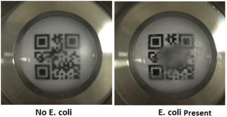
As seen in the figure above when E. coli are present, the droplets clump together and the QR code can’t be read.( Credit: Qifan Zhang)
To precisely quantify the degree of agglutination, the researchers implemented an image processing program to calculate the percentage of area covered by agglutinated Janus emulsions and to evaluate the differences in optical intensity of the images before and after exposure to ConA (concanavalin A, serves as a functional substitute for E. coli bacteria). The program uses the adaptive threshold algorithm to distinguish areas with higher transparency (pristine Janus emulsions) from the opaque regions (agglutinated Janus emulsions).
The MIT team plans to create droplets customized with more complex sugars that would bind to different bacterial proteins. In this paper the researchers used a sugar that binds to E. coli, but they expect that they could adapt the sensor to other pathogens.
The researchers are now working on optimizing the food sample preparation so they can be placed into the wells with the droplets. They also plan to create droplets customized with more complex sugars that would bind to different bacterial proteins. The team leader, Savagatrup says “You could imagine making really selective droplets to catch different bacteria, based on the sugar we put on them”.
The researchers are also trying to improve the sensitivity of the sensor, which currently is similar to existing techniques but has the potential to be much more sensitive, they believe. They hope to launch a company to commercialize the technology within the next year and a half.
Explaining a clear advantage of the technology, one of the lead scientists, Professor Timothy Swager, said: “What we have here is something that can be massively cheaper, with low entry costs. The sensor has been tested out with multiple samples of the infective bacterium and the results are sufficiently successful for the sensor to be considered for commercialization”